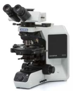
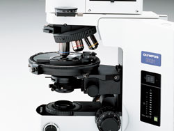
Olympus BX53-P
Clear Observation Images with UIS2 Objective Lens
Thanks to Olympus' sophisticated design and manufacturing technology, new ACHN-P and UPLFLN-P strain-free objectives reduce internal strain to an absolute minimum. Olympus has also totally redesigned its polarizers and polarizing condensers to further enhance performance in polarized light. This means a higher EF* value, resulting in unmatched image brightness and contrast.
*EF value: the ratio of brightness of parallel Nicols to orthogonal(Cross) Nicols. The higher is the EF value, the less is the strain of the optical system. This means that a higher EF value is superior in polarization characteristics.
| Example Observation Images | |
 Quartz Diorite |
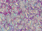 Liquid Crystal MBBA |
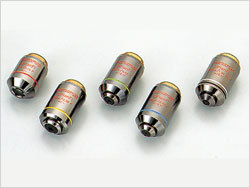 UPLFLN-P Series |
|
|||||||||||||||||||||
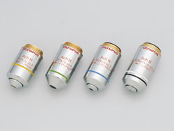 ACHN-P Series |
|
||||||||||||||||||
Superior Operability and Optical Performance
Large Viewfields and Ergonomic Design
Observations can be performed comfortably and efficiently thanks to the Y-shape frame developed in pursuit of ultimate ergonomics. This greatly lessens operator's fatigue during the extended observation.
The Field Number is as large as 22, allowing a 21% wider area than that given by a microscope of ordinary filed number 20 to be observed at a glance. In addition, a bright halogen lamp of 12V-100W is used in the illumination system allowing observation of clear images of polarization.
Rugged and Accurate Rotating Stage
The rotating-centering mechanism attached to the rotary stage allows smooth rotation of a specimen. In addition, there is a click-stop mechanism provided at each 45 degrees for precise measurement. With the option of adding a dual-mechanical stage further discreet x-y movement is possible.
 Rotary Stage |
 Dual-mechanical Stage |
Accessories Suited for High-level Polarization Observation
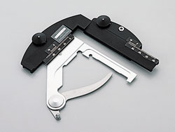
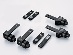
Compensators
 WavePlate/Compensator |
Six different compensators are available for the BX51-P microscope, allowing measurement of various retardation levels, ranging from 0 to 20λ. For easier measurement, the direct readout method is featured. Higher image contrast can be attained by using a Senarmont or Brace- Koehler compensator to change the retardation level in the entire field of view. |
| Retardation Measuring range of compensators | ||
| Compensator | Measuring Range | Major Application |
| U-CTB Thick Berek | 0-11,000nm | Large Retardation Measurement (R*>3λ), ( Crystal , Giant Molecule, Fiber, PhotoelasticStrain, etc.) |
| U-CBE Berek | 0-1,640nm | Retardation Measurement ( Crystal , Giant Molecule, Fiber, Body Tissue, etc.) |
| U-CSE Senarmont | 0-546nm | Retardation Measurement ( Crystal , Body Tissue, etc.) Contrast Intensification (Body Tissue, etc.) |
| U-CBR1 Brace-Kohler 1/10λ | 0-55nm | Minute Retardation Measurement ( Crystal , Body Tissue, etc.) Contrast Intensification (Body Tissue, etc.) |
| U-CBR2 Brace-Kohler 1/30λ | 0-20nm | |
| U-CWE2 Quarts Wedge | 500-2,200nm | Preliminary Survey of Retardation ( Crystal , Giant Molecule, etc.) |
* R stands for retardation.
The use of an interference filter 45IF546 together is recommended for improving measuring accuracy (Except for U-CWE2).
Unmatched image sharpness in orthoscopic and conoscopic observations.
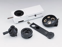 Conoscopic Orthoscope Observation Units |
With a U-CPA conoscopic observation attachment, changeover between orthoscopic and conoscopic observation is simple and quick. Focusing of conoscopic images is easy and accurate. Employing a Bertrand field stop makes it possible to obtain consistently sharp and clear conoscopic images |
Easy adaptation of digital cameras, image analysis software and automated accessories
Our full line of digital cameras provide viewing and speedy image transfer to computers. OLYMPUS Image Analysis Software has been designed specifically for industrial microscopy applications with intuitive menus and advanced software routines. Users are empowered with the latest image analysis and management solutions to satisfy specific application requirements.